Case selection for tooth 23
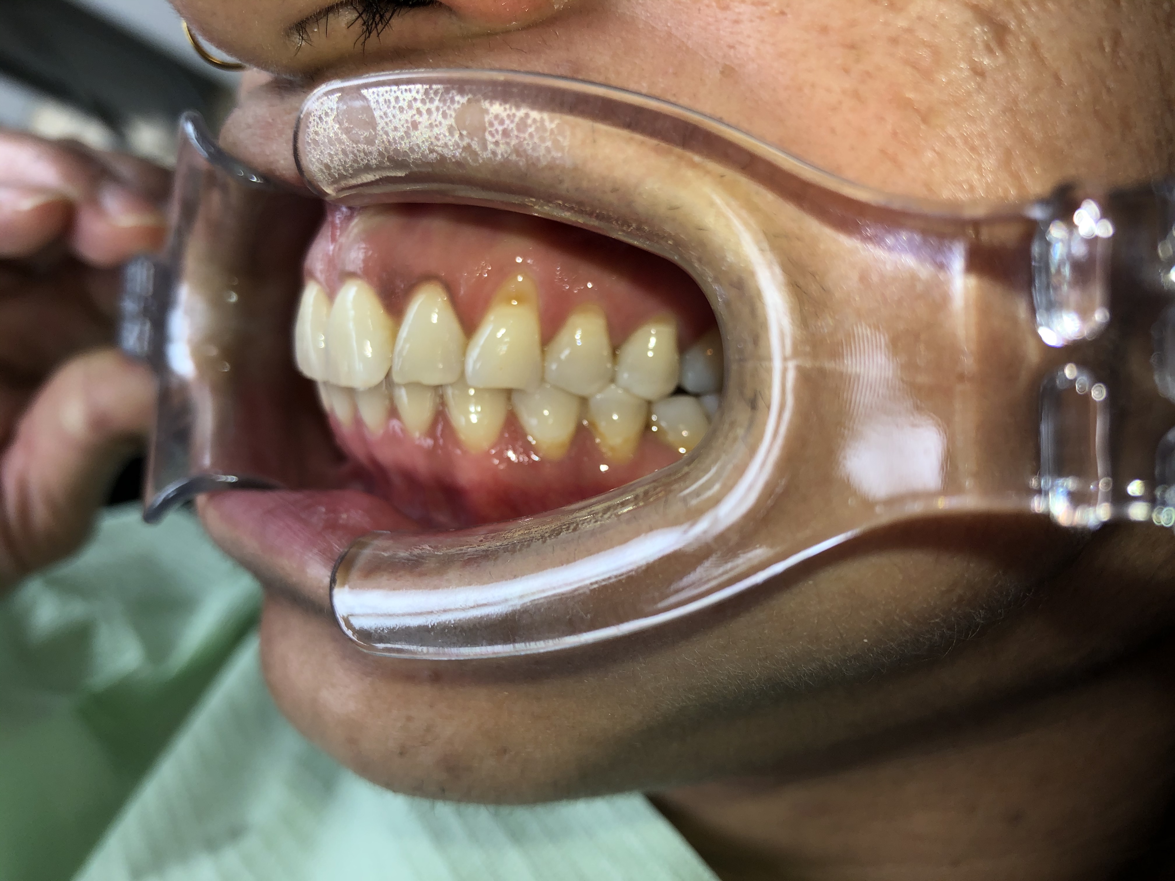
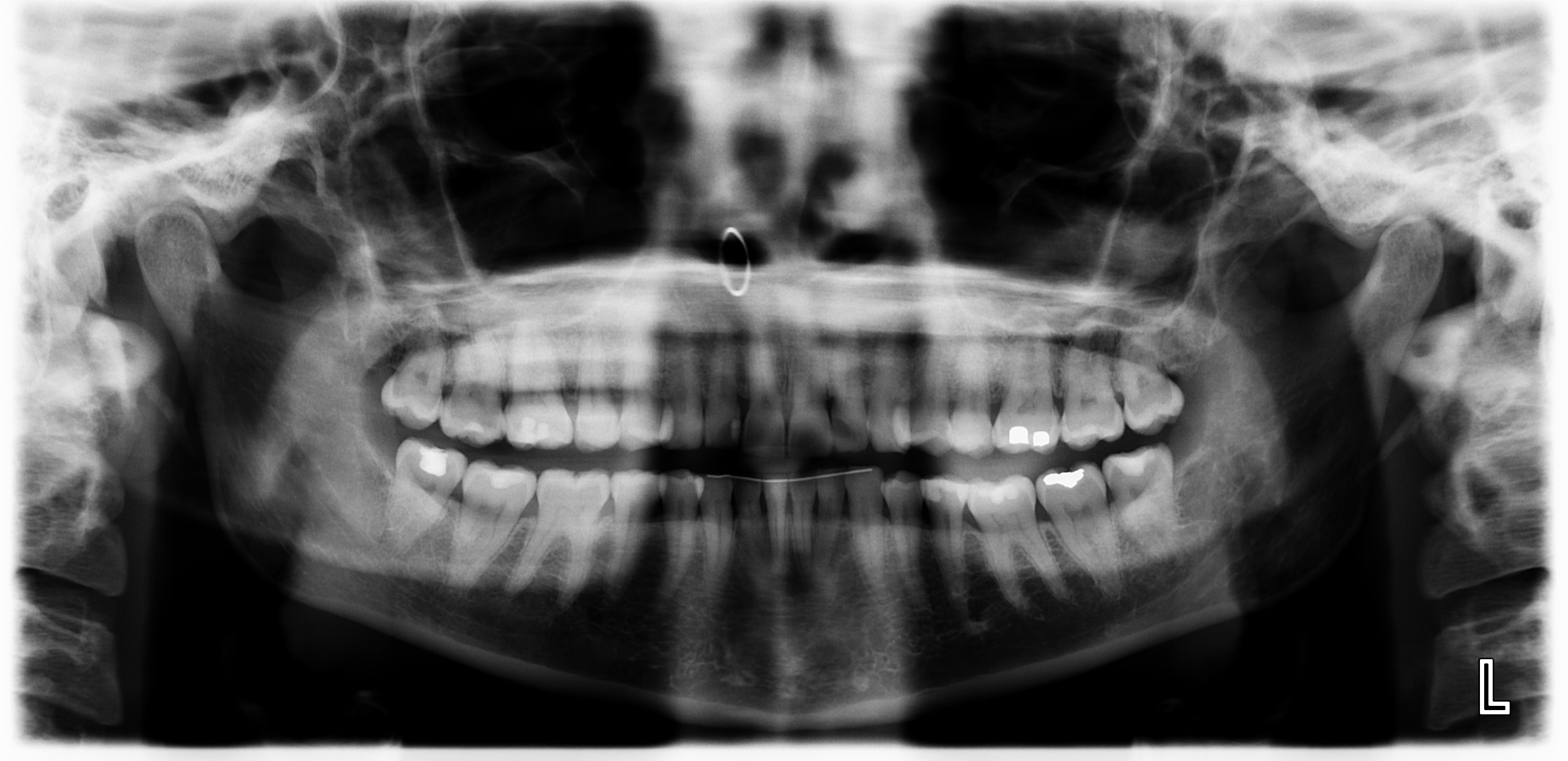
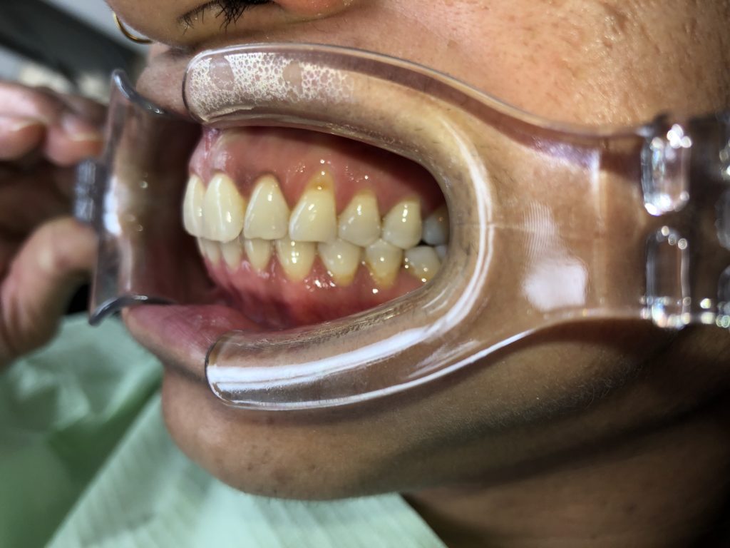
Hi, looking for opinions and advice on this case. Patient is female, 36yo, allergy to sulpha, is on Synthroid 125mg/day, is on a 6 month hygiene program with the office with good oral hygiene. Her cheif concern is the darkness above the left canine. I’ve included intraoral images, a pan and a periapical in the left lateral position. I understand it’s not an accurate image of the upper left canine but it’s what is currently available. Would this be a good candidate for a combination buccal restoration and gingival graft? Looking forward to hearing responses.


in this case we have two issue to consider: 1) loss of enamel and dentin at the cervical margin to due to non-carious cervical lesion: this can cause uncertainty as to where the CEJ is. 2) There has been some horizontal bone loss due to chronic periodontitis which will limit the amount of root coverage. In this case you will get limited root coverage and an improved tissue biotype with soft tissue grafting. Start with a composition restoration up to the anticipate line of maximum root coverage, which in this case will be 1mm apical to the CEJ. Bevel slightly the edges of the crown and root potion of the cervical lesion to reduce the depth of the cervical lesion and to avoid a composite bulge at the transition between the apical portion of the composite and the marginal gingival tissues. Place the composite and finish it supragingivally, then perform the soft tissue grafting with dermal matrix. with the coronally repositioned flap resting on the composite for maximum stability. Patient should be informed of root coverage limitation due to pre-existing horizontal bone loss.